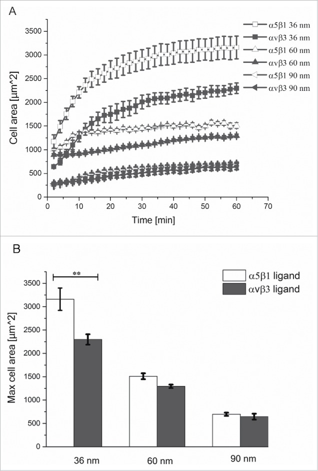
Figure 1.

Cell spreading kinetics on nanopatterned surfaces functionalized with integrin selective ligands. (A) Progression of projected cell area during spreading on nanopatterned surfaces with interparticle distances of 30, 60, or 90 nm, and functionalized with α5β1 (white) and αvβ3 (black) integrin selective ligands. (B) Maximum projected cell area on the different surfaces. Error bars indicate SEM of 3 independent repeats.
