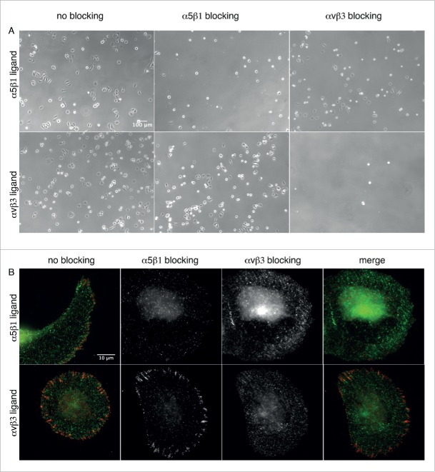
Figure 4.
α5β1 and αvβ3 integrin blocking. (A) Phase contrast micrographs of U2OS cells incubated with α5β1 and αvβ3 integrin selective ligands, and seeded on nanopatterned surfaces functionalized with these ligands. Upper row: Cells adhering to α5β1integrin selective ligands. Lower row: Cells adhering to αvβ3 integrin selective ligands. Left: No integrin blocking. Middle: α5β1 integrin blocking. Right: αvβ3 integrin blocking. (B) Indirect immunofluorescence staining of α5 (green) and αvβ3 clusters (red) in U2OS cells pre-incubated with the integrin selective ligands. Cells were seen to adhere to nanopatterned surfaces functionalized with α5β1 (upper row) and αvβ3 integrin selective ligands (lower row).

