
Figure 5.
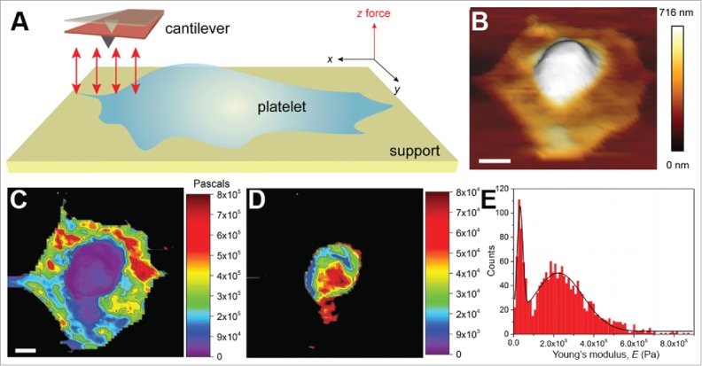
Mechanical investigation of platelets. (A) Schematic illustrating the principle of mechanical characterization of platelets. A platelet is allowed to spread on a fibrinogen-coated glass surface in a physiological buffer (e.g., Tyrode's buffer containing 1 mM each of Ca2+ and Mg2+ ions, ± thrombin). Unlike the conventional AFM contact mode imaging, here F-D curves are recorded (red arrows) in the x-y plane. The F-D curves are used to extract the desired material properties. (B) A 3D topograph of a platelet spread on a fibrinogen-coated surface. The platelet shows a typical fried-egg shape, a high central region and flat periphery, indicative of an adhering platelet. (C) F-D curves were acquired by indenting the platelet with a force of 500 pN at an approach-retract velocity of 5 µm/s. The extension curves were then fit to the Hertz model to obtain the Young's moduli, contour-mapped on the platelet surface. The central region was observed to be softer than the peripheral region. The stiff glass background is shown in black. (D) A resolution of ~137 nm (64 × 64 pixel map of 8.74 × 8.74 µm2) was reasonable to obtain a contour map specifically of the central region. (E) A histogram of the modulus values, 32 kPa for the central region and ~224 kPa for the peripheral region. Scale bars (B, C): 1 µm. C and D are the same scale.