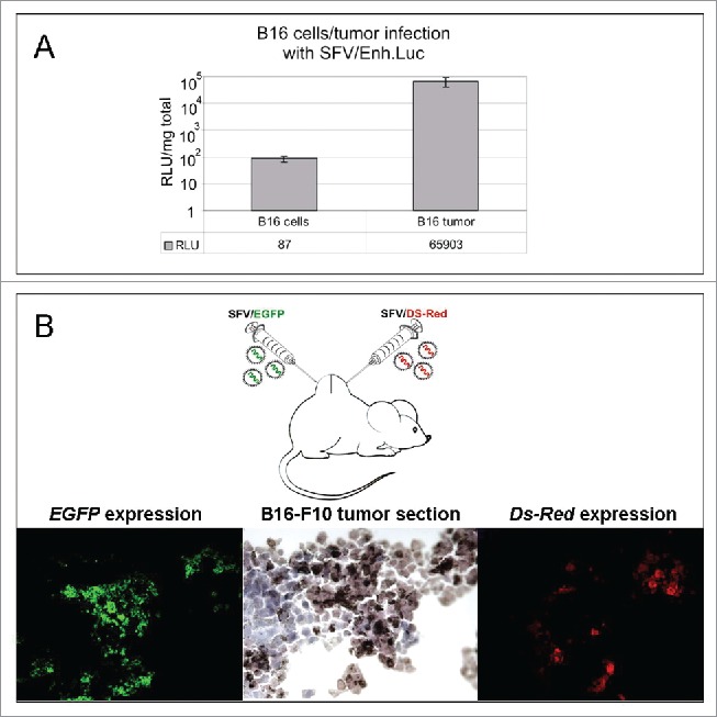
Figure 1.

SFV expression and intratumoral spread in a melanoma mouse model. (A) Infection of B16 melanoma cells in vitro and B16 tumor cells in vivo with SFV/Enh.Luc vector. The B16 cells were infected with SFV at an MOI of 10 in vitro. For the in vivo experiment, B16 tumor-bearing mice were i.t. inoculated with 108 SFV v.p. The luciferase expression analysis in cell lysates and tumor homogenates was performed 24 h post-infection by luminometry. The bar graph presents the RLUs per 1 mg protein in the cell lysate/tumor homogenate. The results represent the mean ± s.e. RLU - relative light unit. (B) Administration strategy of SFV vectors and fluorescence microscopy of B16 tumor cryosections, demonstrating SFV/FGFP and SFV/Ds-Red virus spread in the tumor. A total of 106 v.p. of SFV/EGFP and SFV/Ds-Red were injected in different tumor sides by direct intratumoral injections. The tumors were cryosectioned and analyzed 24 h after SFV vector administration.
