
Figure 5.
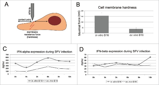
Comparison of cell membrane elasticity and IFN-α/β expression between in vitro and first-passage ex vivo B16 cells. The principle of AFM measurement is schematically demonstrated in panel A. The middle point between the cell nucleus and the elongated cell body was selected as the contact point of the AFM tip and the cell surface. The pushing force was increased slowly until the membrane was punctured. The acquired maximal forces essential for membrane puncturing of in vitro and ex vivo B16 cells are shown in panel B. The membrane resistance force indicates the elasticity of the sample surface; higher maximal force indicates higher membrane hardness. The results represent the mean ± s.e. Expression levels of IFN-α (C) and IFN-β (D) were determined in in vitro and first-passage ex vivo B16 cell lysates before SFV infection (0 h) and at 1 h, 3 h, 6 h, 9 h and 18 h post-infection. The results represent the concentration of IFN protein per 1 ml of cell lysate (pg/ml).