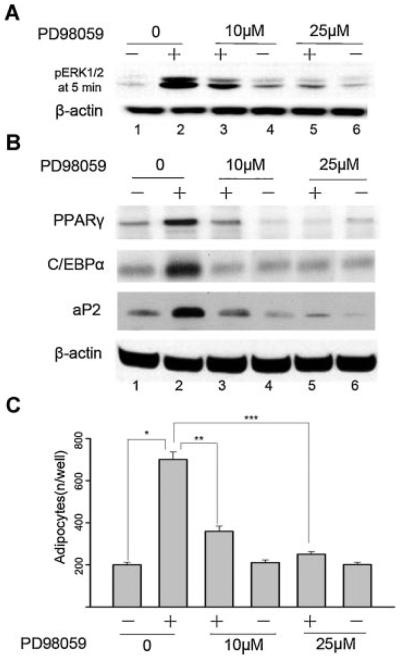
Fig. 2.
Effect of blockade of ERK1/2 activity on adipogenic differentiation of mouse BMSC cultures. Cells were cultured in control medium (−) or adipogenic cocktail (+) or adipogenic cocktail supplemented with PD98059. A: Western blotting assay for pERK1/2 was performed at day 5 after 5 min exposure to the adipogenic cocktail. B: Western blotting assay for PPARγ, C/EBPα, and aP2 were performed at day 21 as described under “Materials and Methods Section.” Anti-β-actin antibody was used as a control. Results are representative of three independent experiments. C: The number of Oil Red O-positive adipocytes was calculated at day 21. The data are mean ± SD. *P < 0.05 versus non-treated vehicle. **P < 0.05 versus adipogenic cocktail-treated alone. ***P < 0.05 versus adipogenic cocktail-treated alone. Results are representative of three independent experiments.

