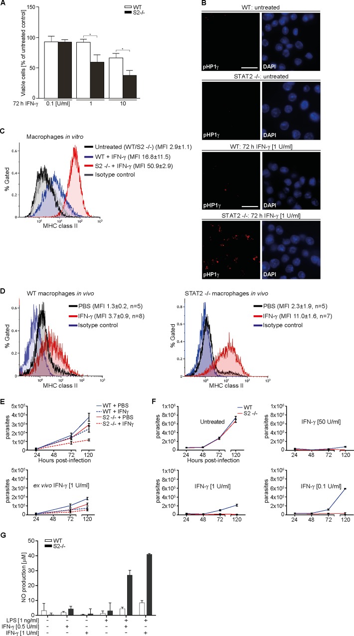
Fig 6. STAT2 modulates key IFN-γ effector functions.
(A) Viability of immortalized macrophages was measured by Alamar blue assay of mitochondrial function after 72 h in the presence of IFN-γ. (B) Widefield microscopy detecting HP1γ (anti-phospho-S83 HP1γ antibody) in immortalized macrophages before (top four panels) and after (bottom four panels) treatment with IFN-γ. Nuclei were DAPI stained. Scale bar = 15 μm. (C) Major histocompatibility complex (MHC) II expression on immortalized WT and STAT2-/- macrophages treated without or with 0.1 U/ml IFN-γ for 72 h was determined by fluorescence-assisted cell sorting (FACS) using Alexa Fluor 488-conjugated specific and isotype control antibodies. Gating for live cells was performed using forward and side scatter analysis, and the same gate was used for the histograms in (C) and (D). MFI, median fluorescence intensity. (D) Same as (C), but using peritoneal leukocytes from mice injected with PBS or IFN-γ. MHC class II expression (PerCP-conjugated antibody) was analyzed on macrophages detected by Alexa-Fluor 488-conjugated anti-F4/80 antibody. A representative histogram is shown and median PerCP fluorescence intensities ± s.d. per cohort (n = animal numbers) are given. (E) Peritoneal macrophages from PBS or IFN-γ-injected mice were cultured without (top) or with added IFN-γ (bottom) in duplicates, infected with Toxoplasma gondii, and extracellular parasites were counted at the indicated time points. Graphs combine the results from five control and eight experimental mice per genotype, bars show mean and s.d. (F) T. gondii release from infected immortalized macrophages treated with different IFN-γ concentrations. Results represent two independent experiments, bars show mean and s.d. (G) Nitric oxide (NO) production by immortalized macrophages determined using Griess reagent in triplicates after 36 h of the indicated treatments, bars show mean and s.d. See S1 Data for raw data.

