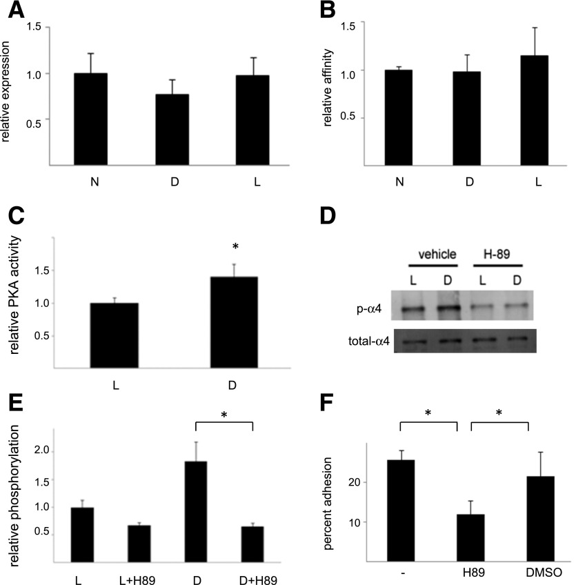
Figure 2.
Growth in high glucose simulates PKA activity, α4-integrin phosphorylation, and PKA-dependent adhesion. A: α4 expression. ECFCs were grown for 1 week in normal media (N), in media containing 25 mmol/L d-glucose (D), or in the osmotic control media (L), and α4-integrin expression was determined by flow cytometry. n = 4. B: α4β1 affinity. The same cells were used to determine α4β1 affinity by flow cytometry. n = 3. C: PKA activity. ECFCs were grown in media containing 25 mmol/L d-glucose and the osmotic control media for 1 week, and PKA activity was then determined. n = 10. *P < 0.05. D: α4 phosphorylation. Cells grown for 1 week in the same media as indicated were treated with DMSO (vehicle) or the PKA inhibitor H89 and levels of phospho-α4 were determined by Western blotting. Illustrated is a representative blot. E: Illustrated are group data for high glucose–stimulated α4 phosphorylation. n = 8. *P < 0.05. F: Cell adhesion. Cells grown in d-glucose media were left untreated or were treated with either H89 or DMSO, and then adhesion to an S17 cell monolayer was determined after 30 min. n = 4. *P < 0.05.

