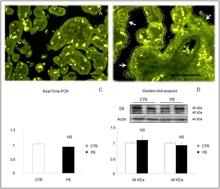
Fig 1. Representative images of immunofluorescent staining for D6 in placental sections from (A) a normal pregnant woman at term or (B) a woman with PE at 28 weeks of gestation.
Characteristic positive expression for D6 receptor was detected in the syncytiotrophoblast monolayer in PE (B). Scale bar 100 μm. No significant differences were found between PE and control placental lysates in terms of overall D6 tissue expression by RT-PCR (C) or Western blot analysis (D). Results are expressed as mean ± SE of six experiments. NS: not significant.

