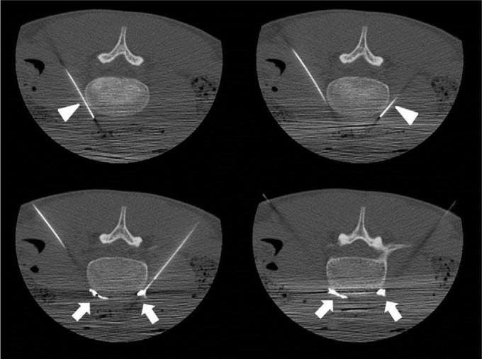
Figure 4.

Axial CT fluoroscopy images patient 2
Notes: Axial CT fluoroscopy images obtained during LSB showing sequential placement of 21 G, 15 cm needles (top left and top right, arrowheads) into the prevertebral space at the L4 vertebral level. Images obtained following administration of medication and contrast through each needle show even dispersion of the agents in the prevertebral space adjacent to the lumbar sympathetic chains (bottom left and bottom right, arrows).
Abbreviations: CT, computed tomography; LSB, lumbar sympathetic blockade.
