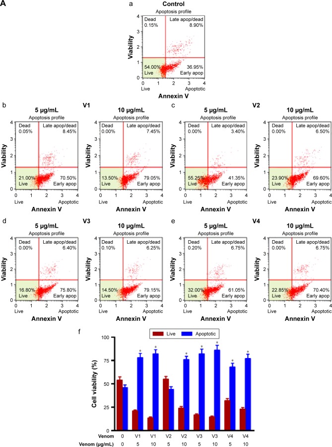
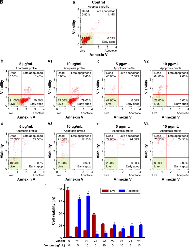
Figure 5.
Assessment of apoptosis in (A) HCT-8 and (B) MDA-MB-231 cell lines treated with different snake venoms for 24 hours.
Notes: The cells cultured in either DMEM or RPMI medium in the absence of venoms were used as control. Extent of apoptosis was detected by Annexin V–PI dual staining. The percentage of total apoptotic cells (early + late) was calculated and shown in the bar graphs (f), a=control, b= v1; 5 µg/ml and 10 µg/ml c= v2; 5 µg/ml and 10 µg/ml, d= v3; 5 µg/ml and 10 µg/ml, e= v4; 5 µg/ml and 10 µg/ml. The number of apoptotic cells was higher in the venom-treated cell lines when compared with the control group. *Statistically significant (P<0.05). V1, V2, V3, and V4 are the venoms obtained from the species of the snakes, namely Bitis arietans, Cerastes gasperettii, Echis coloratus, and Echis pyramidum, respectively.
Abbreviations: DMEM, Dulbecco’s Modified Eagle’s Medium; PI, propidium iodine; RPMI, Roswell Park Memorial Institute.


