Figure 1.
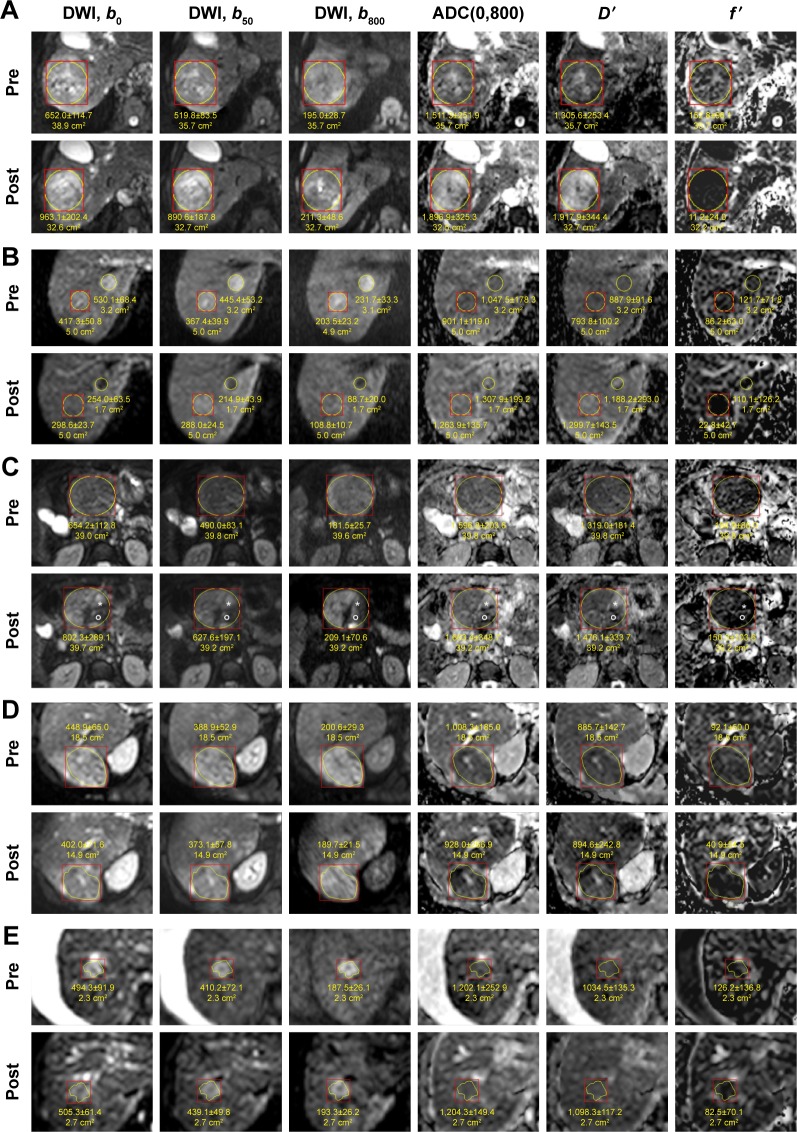
Diffusion-weighted images with maps of ADC(0,800), D′, and f′ pre- and post-TACE (A–C, E) and RE (D).
Notes: Examples with different changes in LDT and LDCE can be seen: (A) HCC with a decrease of 24% in LDT and 100% in LDCE (Group A, responders). The homogeneous increase in ADC(0,800) might indicate necrosis over the entire tumor area. This is supported by the stronger increase in D′ accompanied by the decrease in f′. The degree of necrosis might be underestimated by ADC(0,800). (B) The decrease in LDT and LDCE was 10% and 100%, respectively, for both lesions (Group A). After therapy, both HCCs show an increase in ACD(0,800), and they appear isointense in the postimage but were slightly hypointense in the preimage. In the DWI images, both HCCs appeared hypointense after therapy, but also in the b0 image. This might be due to coagulative necrosis and is supported by the even stronger increase in D′ and the decrease in f′. (C) This HCC with a decrease of 5% in LDT and 67% in LDCE (Group A) shows heterogeneous response. The main part remains hyperintense in the b800 postimage with no changes in ADC(0,800), D′ and f′ indicating vital tumor. A small area (star) shows clear increase in ADC(0,800) and D′ and decrease in f′, which are in accordance with necrosis. Another small area (round circle), which appears hypointense in all the DWI images, shows no increase in ADC(0,800) and D′, but a decrease in f′. This might indicate coagulative necrosis or hemorrhage or embolization. (D) HCC with a decrease of 10% in LDT and 27% in LDCE (Group A). The intensity on contrast-enhanced images (not shown) is also decreased. On DWI, the area of hyperintensity is decreased, but the ADC(0,800) is slightly decreased. However, D′ and f′ show that the decrease in ADC(0,800) is caused by a decrease in f′, whereas D′ is rather unchanged. This might be associated with embolization or necrosis of low degree. (E) HCC with an increase of 11% in LDT and 19% in LDCE (Group B, nonresponders). On DWI, the area of hyperintensity is also slightly increased. ADC(0,800) and D′ values are rather unchanged, indicating vital tumor. The slight decrease of f′ may be caused by embolization.
Abbreviations: ADC, apparent diffusion coefficient; DWI, diffusion-weighted imaging; HCC, hepatocellular carcinoma; LDCE, longest diameter of the region with contrast enhancement; LDT, longest diameter of the whole tumor on morphological images; RE, radioembolization; TACE, transcatheter arterial chemoembolization.

