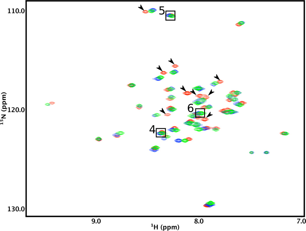
Figure 4.
Comparison of the Lmod2s1 NMR spectra in the presence of Tpm1.1 model peptides of varying lengths. The 2D 15N-HSQC spectra of 15N-labeled Lmod2s1 were recorded in the presence of a >20% stoichiometric excess of αTM1a1-14Zip (red), αTM1a1-21Zip (green), or αTM1a1-28Zip (blue). Arrows show the cross-peaks of the Lmod2s1/αTM1a1-14Zip complex that are markedly shifted with respect to those of the Lmod2s1/αTM1a1-28Zip complex. The cross-peaks of residues 4–6 are not affected by binding and are shown in boxes.

