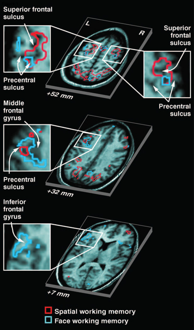
Figure 1.
(A) Areas in PFC that showed elevated delay period activation as measured with fMRI during memory for either face identity or a spatial location. Partially segregated areas were observed that responded more during STM for one type of content versus the other (From Courtney et al., 1998). Reprinted with permission from the authors and the original publisher.

