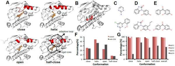
Figure 2. HSP90 adopts at least four ligand-induced binding modes.
A. Four conformations of HSP90 ligand-induced binding pocket based on the nearby adaptive loop (L2, between H4 and H5 [43]): close (2WI5), helix (4EFU), open (3RLR), half-close (3B28) (white cartoon: HSP90, red cartoon: flexible loop, orange ticks: small molecules) B. Four waters in the binding pockets labeled from 1 to 4 (white cartoon: HSP90, red sphere: water molecules) C. Aminopyrimidine scaffold and compound (2XDX) D. Benzimidazolone scaffold and compound (4YKR) E. Benzophenone-like scaffold and compound (4YKR) F. Histogram of binding modes among the N=181 known co-crystal structures and I=69 structures with IC50 data. (N: number of co-crystals, I: number of co-crystal with IC50 data) G. Histogram of conservation frequency of water molecule in Fig. 2B shows that three crystal waters are 100% conserved.

