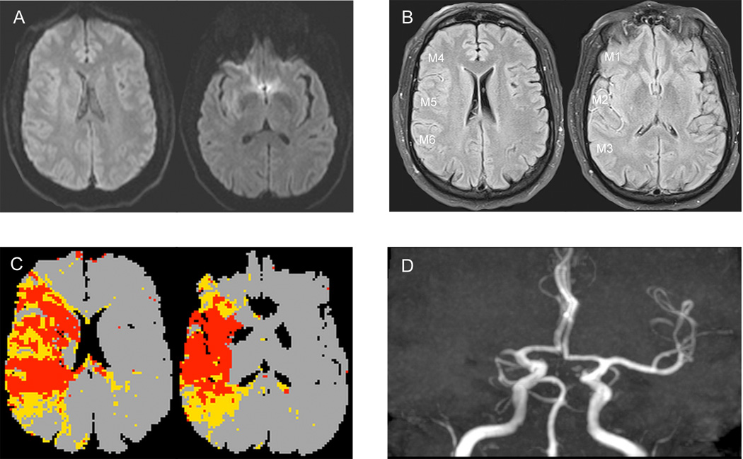
Figure 1.
Illustrative case of FVH-ASPECTS evaluation in a patient with a right MCA occlusion (A–D). No hyperintense lesions are visible in the right MCA territory (A). FVH-T-ASPECT score was 11 (M1=1, M2=2, M3=2, M4=2, M5=2, M6=2), FVH-O-ASPECTS was 11 and FVH-I-ASPECTS was 0 (B). PWI showed that mismatch on the Tmax map was congruent with the FVHs distribution(C).
Abbreviations: FVH-T-ASPECTS=total FVH-ASPECT score, FVH-O-ASPECTS=FVH-ASPECT score outside DWI-positive area, FVH-I-ASPECTS=FVH-ASPECT score inside DWI-positive area

