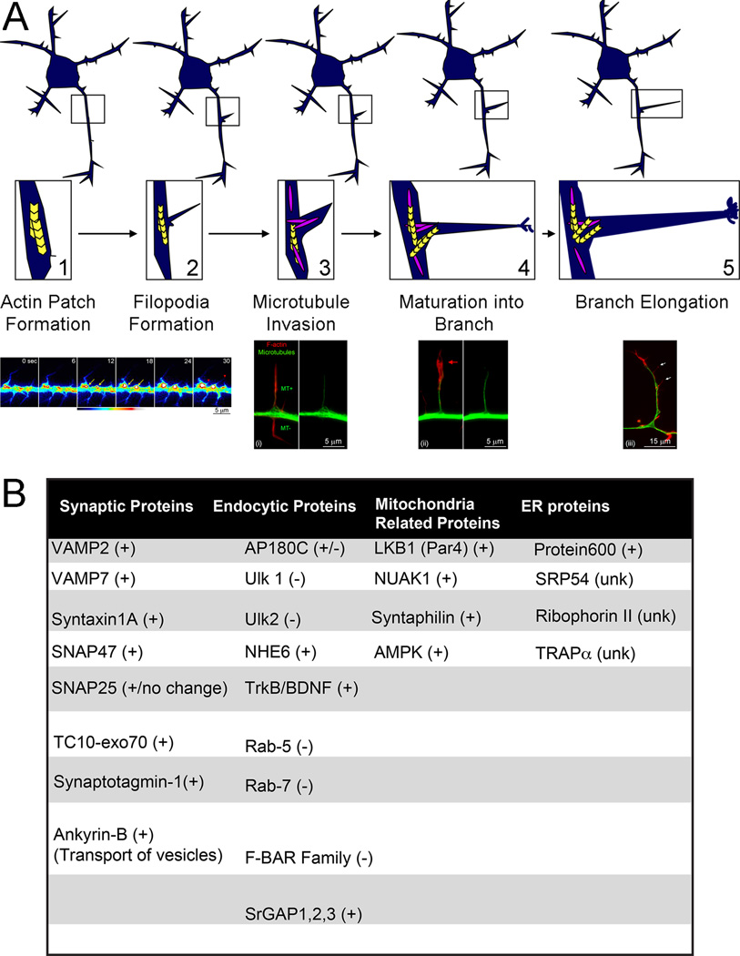
Figure 1.
Sequence of cytoskeletal events underlying the collateral branching of axons. (A) Schematic of the sequence of events leading to the formation of a collateral branch (top, A1–A5). Examples of cytoskeletal dynamics and organization during the branching of embryonic sensory axons are also provided (bottom). Axonal filopodia are the first morphogenetic step in the formation of a branch (A2). Filopodia emerge from pre-existing patches of axonal actin filaments (A1). The time-lapse sequence (A1 bottom) shows the formation of axonal actin patches and the emergence of filopodia from the patches (eYFP-β-actin imaging; time is shown in seconds in each panel). Actin patches form and elaborate (i.e., grow brighter and larger) as denoted by the yellow arrows at 12–18 sec. The red arrowheads and 30 sec denote filopodia that have emerged from the two patches labeled in prior panels. The targeting of one or more microtubules (MT+) into a filopodium is a required event for the maturation of the filopodium into a branch (A3). Filopodia that are targeted by microtubules can then mature into a branch (A4). The maturation of the filopodium is characterized by the development of a polarized distribution of actin filaments (red arrow, A4 bottom), which form a small growth cone-like structure at the tip of the nascent branch. As the branch elongates actin filaments remain mostly polarized at its tip, although the branch can also give rise to filopodia along its shaft (white arrows, A5 bottom), and in turn secondary branches. (B) Table featuring the proteins discussed in the text and denoting whether they promote (+) or impede (−) branching. (unk) denotes proteins with insufficient data regarding their role in axon branching.

