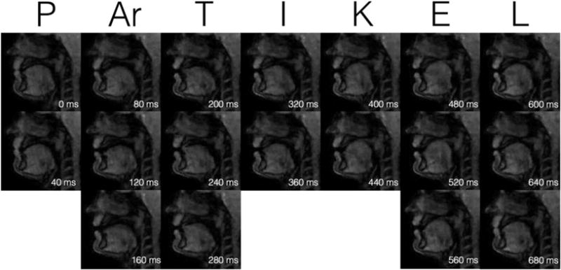
FIGURE 10.

Example of acquisition with Protocol 3: The raw data were acquired with a radial gradient echo sequence with golden angle rotation. Images were reconstructed using a conjugate gradient-SENSE method that applies regularization using a spatial total variation (TV) operator. Twenty-five subsequent echoes were used to calculate low resolution sensitivity maps for each coil and a complete image with a spatial resolution of 1.8 × 1.8 × 10 mm3, leading to a native temporal resolution of 55 msec that was further accelerated to 40 msec by applying a sliding window. (Figure courtesy: M. Burdumy, University Medical Center Freiburg, Germany.)
