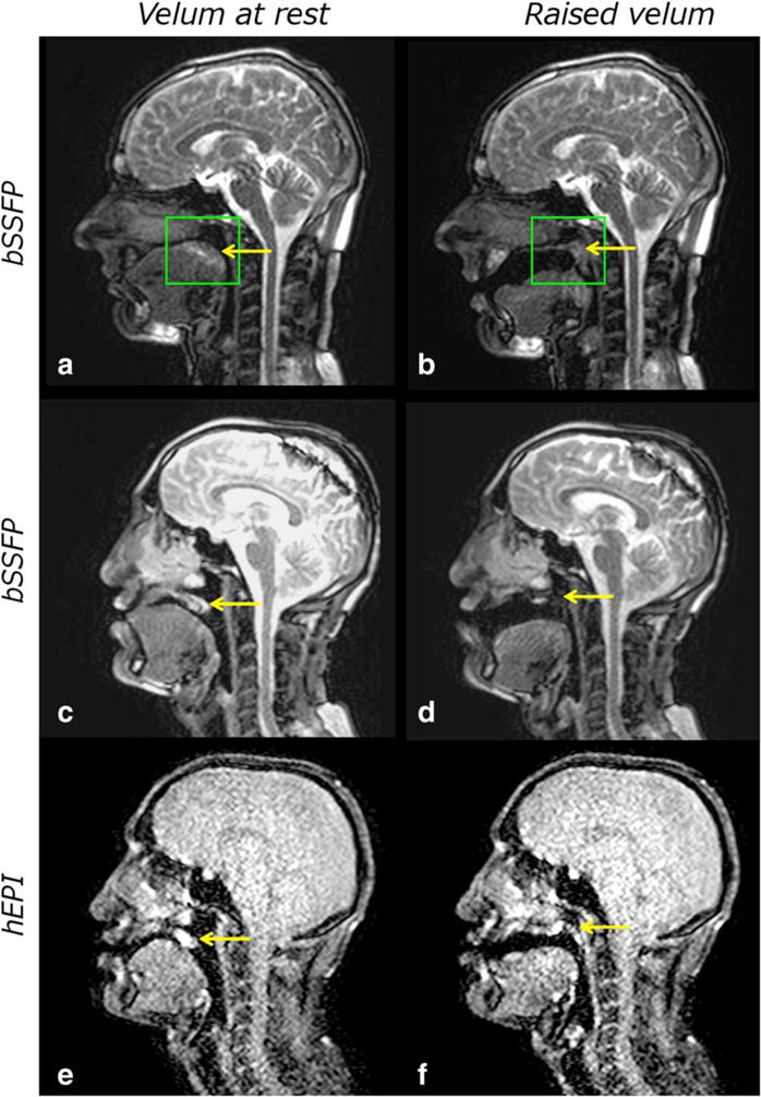
FIGURE 4.

Example images of velopharyngeal closure obtained at 1.5T using protocol 1. (a,b) The placement of the shim volume (green box) centered around the velum (yellow arrow) for a 39-year-old male volunteer. Despite careful shimming images can degrade substantially in some subjects during the speech sample acquisition as demonstrated by images (c,d) in a 28-year-old patient with repaired cleft lip and palate. The velum (yellow arrow) almost completely disappears in the elevated position (d). Although h-EPI sequences have a lower SNR (e,f), it is possible to track the velum (yellow arrow) throughout the speech sample.
