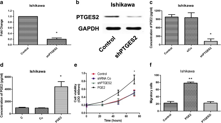
Fig. 2.
Prostaglandin E2 raises proliferation and invasion in human endometrial cancer cells a RT-PCR analysis for Ishikawa cells after transfection of PTGES2 shRNAs. *p < 0.05 versus control group, tested with unpaired Student’s t test. b Western blot tests for Ishikawa cells after transfection of PTGES2 shRNAs. c ELISA for PGE2 concentration in shRNAs transfected Ishikawa cells after 24 h of culture. *p < 0.05, analyzed by one-way analysis of variance (ANOVA). d ELISA for PGE2 concentration in PGE2 stimulated Ishikawa cells after 24 h of culture. C replicated control group, in which no PGE2 stimulated. C0 replicated PGE2 working concentration. *p < 0.05, analyzed by one-way analysis of variance (ANOVA). e CCK8 assays were conducted at each time point to quantify cell viability for Ishikawa cells transfected with control or PTGES2 shRNA, or stimulated with PGE2. *p < 0.05, analyzed by one-way analysis of variance (ANOVA). f Transwell for Ishikawa cells with PGE2 stimulated or transfected with PTGES2 shRNA. Figure shows the number of invasive cells for each group (averaged across five random images). **p < 0.01, analyzed by one-way analysis of variance (ANOVA)

