
FIGURE 7.
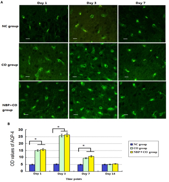
The OD value alterations of AQP-4 expression in NC, CO, and NBP+CO groups. A small amount of AQP-4 positive cells scattered in brain tissue in normal rats. After exposure to CO, AQP-4 expression was temporarily elevated and then gradually decreased. NBP treatment caused a slightly higher expression of AQP-4 protein compared to that in CO group, whereas it lacked a statistically significant difference between the two groups (n = 4, P > 0.05). (A) The expressions of AQP-4 positive cells in each group; (B) The OD value alterations of AQP-4 positive cells in different groups (n = 4). ∗Compared with NC group (n = 4, P < 0.05 (F = 10.621∼56.349). Scale bar = 30 μm.