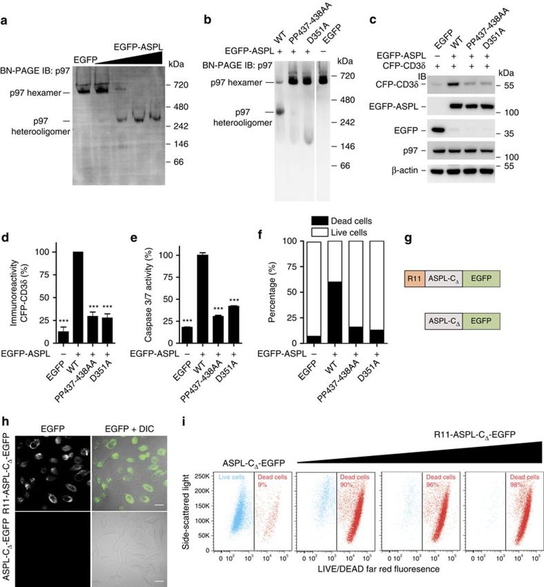
Figure 5. ASPL-induced disruption of p97 hexamers causes mammalian cell death.
(a) Overproduction of EGFP-ASPL causes concentration-dependent disassembly of endogenous p97 hexamers in HEK293 cells. Protein extracts were analysed by BN-PAGE. p97-containing protein complexes were identified by IB using an anti-p97 antibody. (b) EGFP-ASPL and indicated variants of ASPL were overproduced in HEK293 cells. After 24 h, native protein complexes were resolved by BN-PAGE and immunoblotted to identify p97-containing protein complexes using an anti-p97 antibody. (c) CFP-CD3δ, EGFP-ASPL and indicated variants of ASPL were overproduced in HEK293 cells. After 24 h, total cell lysates were resolved by SDS–PAGE and immunoblotted to detect indicated proteins; antibodies: anti-CD3δ, anti-GFP, anti-p97 and anti-β-actin. (d) The levels of CFP-CD3δ in samples as indicated in c were quantified using an Aida image analyser. Data are expressed as mean±s.e. (n=3). ***P≤0.001 compared with WT-ASPL (Student's t-test). (e) EGFP-ASPL and indicated variants with point mutations were overproduced in HEK293 cells; activation of caspases 3/7 was monitored after 72 h. Data are expressed as mean±s.e.; (n=3). ***P≤0.001 compared with WT-ASPL (Student's t-test). (f) EGFP-ASPL and indicated variants with point mutations were overproduced in HEK293 cells for 72 h; cells were stained with LIVE/DEAD far-red fluorescence stain and live and dead cell populations were quantified using flow cytometry. 10,000 EGFP cells were analysed per each sample. (g) Schematic representation of R11- and EGFP-tagged ASPL-CΔ recombinant proteins. (h) Confocal imaging micrographs of fixed HeLa cells showing the uptake of R11-ASPL-CΔ-EGFP protein into cells. Scale bars, 10 μm. (i) HeLa cells were treated for 24 h with the recombinant proteins R11-ASPL-CΔ-EGFP and ASPL-CΔ-EGFP (control protein), respectively; cells were stained with the LIVE/DEAD far-red fluorescence stain and live and dead cell populations were quantified using flow cytometry.

