Figure 3.
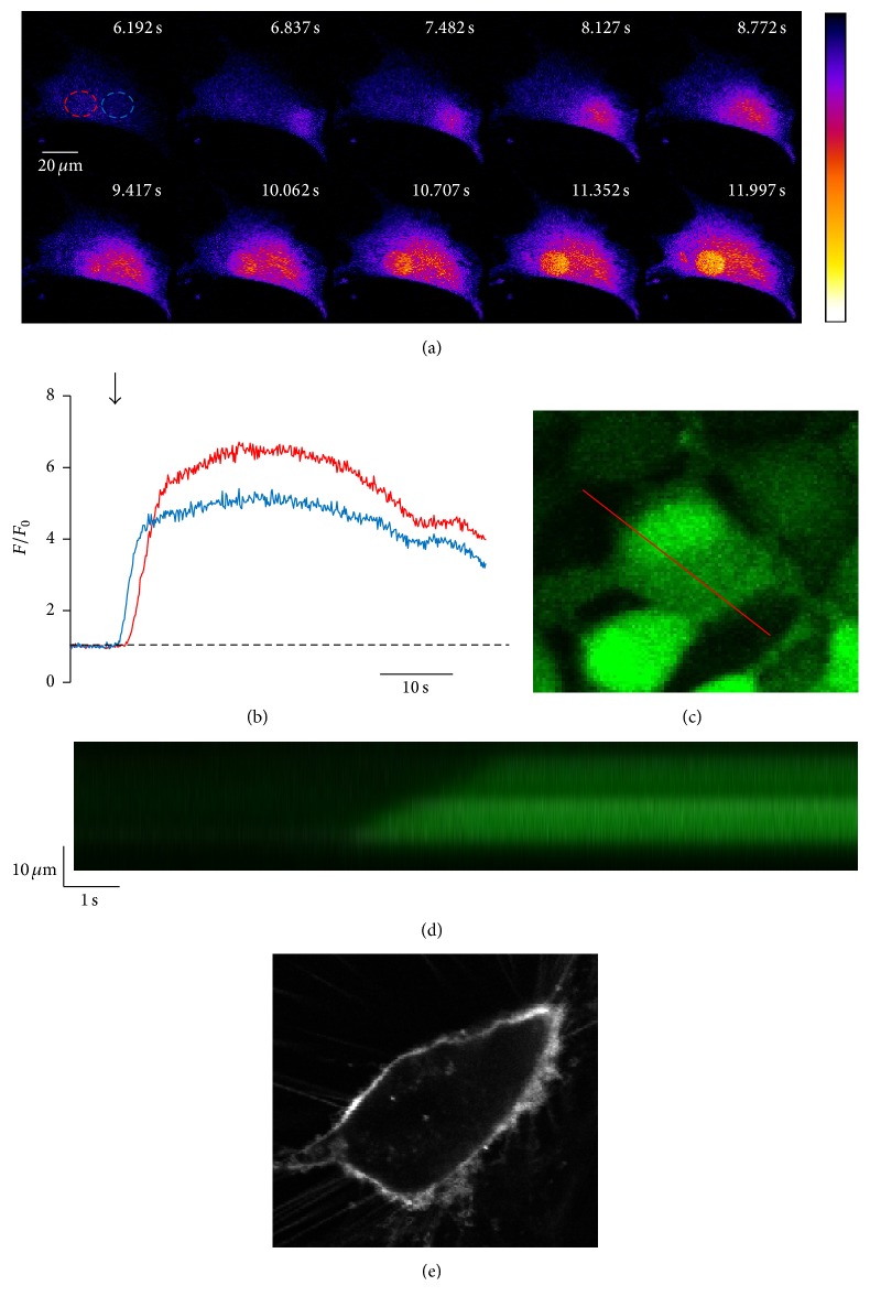
Ca2+ oscillations in electrically stimulated CPCs show spatial heterogeneity and propagate in a wave-like fashion. ((a) and (b)) Representative 2D high-speed confocal image montage of the activation of a Ca2+ oscillation in a CPC loaded with fluo-4/AM. ROIs (red: nuclear, blue: cytosolic) indicated in (a) are graphed in (b). ((c) and (d)) Measured from the red line shown in (c), the confocal line scan in (d) shows that initiation of Ca2+ release at one edge of the cell propagates through the cell to the other side. (e) Representative image of a CPC stained with Di-8-ANEPPS.

