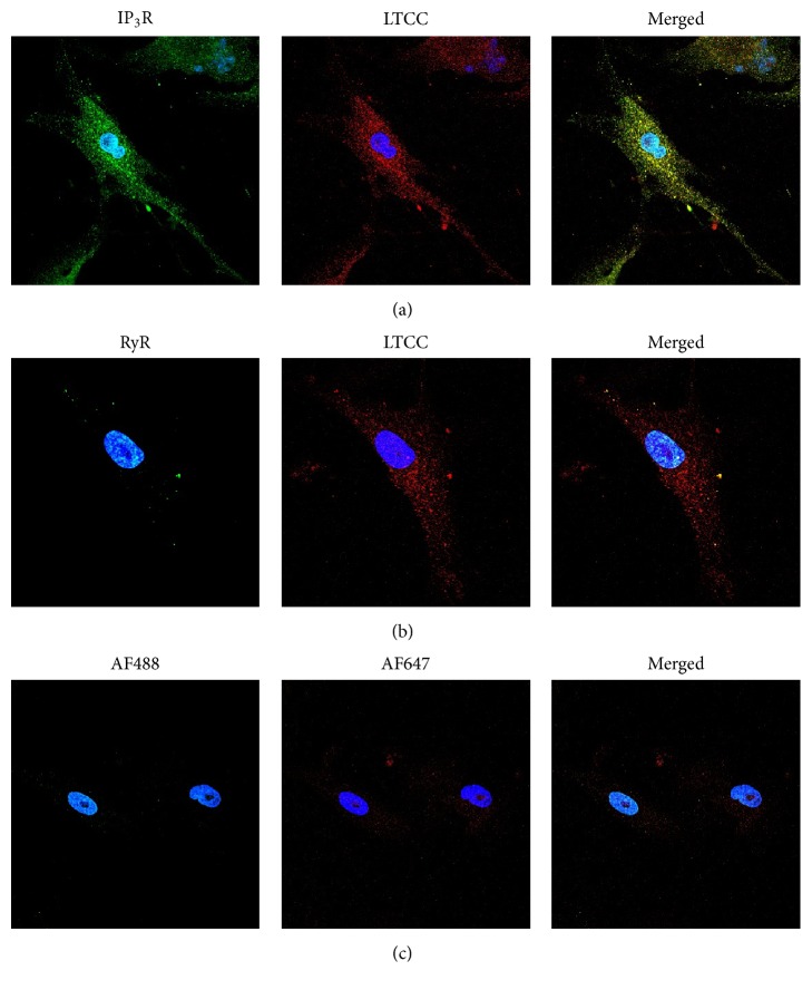
Figure 5.
Immunostaining of CPCs shows expression and localization of the LTCC and IP3R. CPCs were fixed, permeabilized, and incubated with primary antibodies against the L-type Ca2+ channel (LTCC, Cav2.1 subunit), type 2 inositol 1,4,5-trisphosphate receptor (IP3R), or type 2 ryanodine receptor (RyR). Cells were then incubated with either Alexa Fluor 488 (IP3R and RyR; green) or Alexa Fluor 647 (LTCC; red). Nuclei are stained with DAPI (blue). Expression of the IP3R and LTCC is shown in (a), while expression of the LTCC and the RyR is shown in (b). Cells were also incubated with only secondary antibodies to determine nonspecific binding in (c). Images are representative of 3 replicates.

