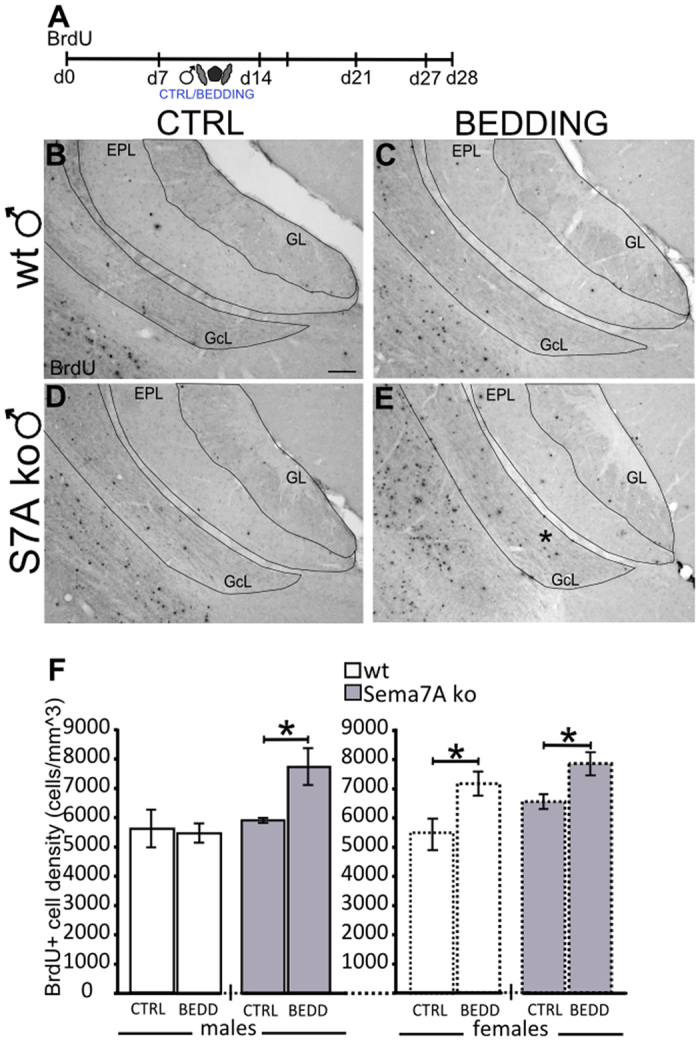
Figure 3. Sema7A ko males display feminized-like neurogenesis in response to male pheromones.

(A) Experimental protocol: newborn cells were quantified in the GcL of the AOB 28 days after BrdU injection. From day 7 to day 14, the mice were familiarized with male-soiled bedding, while the control groups received clean bedding. (B–E) Representative images of AOB sections showing BrdU-positive newborn neurons in wt (B,C) and Sema7A ko (D,E) males after bedding familiarization. (F) Quantification of BrdU-positive cell density in the AOB GcL indicates an increase in newborn neurons in the Sema7A ko males after male-soiled bedding (BEDD) exposure compared to the clean bedding (CTRL) condition (n = 4; unpaired Student’s t-test, P = 0.030); no differences are observed in wt males (n = 4 CTRL, n = 6 BEDD; unpaired Student’s t-test, P > 0.05). Both wt and Sema7A ko females show increased BrdU-positive cell density in the BEDD condition (n = 4 wt, 4 ko) compared to the CTRL condition (n = 4 wt, 5 ko; unpaired Student’s t-test, P = 0.043, P = 0.024, respectively). The values shown are the mean ± s.e.m. Abbreviations: AOB: accessory olfactory bulb; GcL: granule cell layer; GL: glomerular layer; EPL: external plexiform layer; S7A: Semaphorin 7A; BrdU: bromodeoxyuridine. Scale bar: 100 μm.
