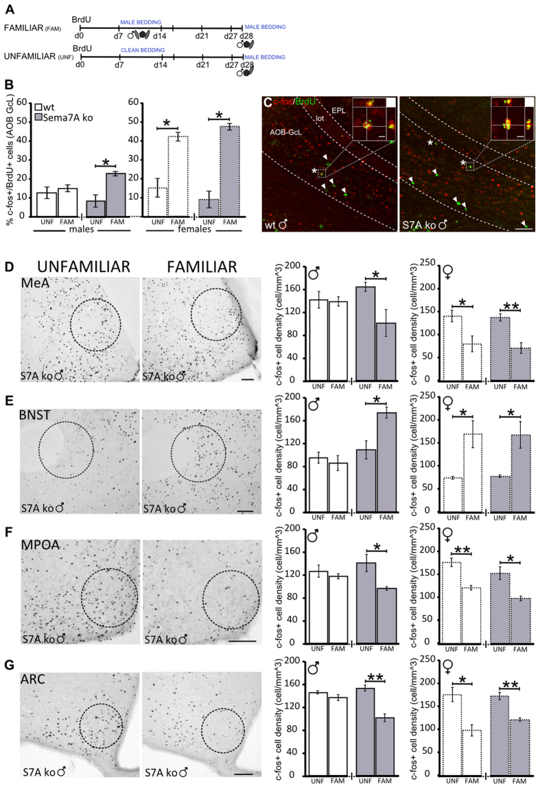
Figure 4. The vomeronasal pathway of Sema7A ko males shows a feminized c-fos response to pheromones.
(A) Experimental protocol: the familiar groups were stimulated with male-soiled bedding during the 2nd week after BrdU administration, from day 7 to day 14, whereas the unfamiliar groups received clean bedding. Both groups were then stimulated with male bedding 90 min before animal perfusion. (B) Quantification of c-fos/BrdU double-labelled cells in the AOB GcL reveals an increase in the familiar Sema7A ko group compared to the unfamiliar Sema7A ko group (n = 4; Wilcoxon-Mann-Withney test, P = 0.028). No differences are observed in wt males (n = 4, Wilcoxon-Mann-Withney test, P > 0.05). An increase in c-fos/BrdU+ cells is also observed in the familiar versus unfamiliar group of wt females (n = 4; Wilcoxon-Mann-Withney test, P = 0.029) and Sema7A ko females (n = 4; Wilcoxon-Mann-Withney test, P = 0.028). (C) BrdU (green) and c-fos (red) immunofluorescence in the AOB-GcL of wt and Sema7A ko males. Asterisks indicate representative c-fos/BrdU double-stained cells. Arrowheads indicate c-fos-negative/BrdU-positive cells. Scale bars: main panels 50 μm; magnified panels 5 μm. (D–G) Representative images of Sema7A ko males and histograms showing changes in c-fos expression throughout the VNS in Sema7A ko and wt mice after bedding familiarization. The quantification of c-fos+ cell density shows no differences between the unfamiliar and familiar groups of wt males (unpaired Student’s t-test, P > 0.05). In Sema7A ko males and in wt and Sema7A ko females, familiarization decreases c-fos cell density in the medial amygdala (MeA, D), medial preoptic area (MPOA, F) and arcuate nucleus (ARC, G) and increases c-fos expression in the bed nucleus of the stria terminalis (BNST, E). (Sema7A ko males n = 3; MeA P = 0.034; BNST P = 0.024; MPOA P = 0.044; ARC P = 0.003; wt females n = 3; MeA P = 0.047; BNST P = 0.033; MPOA P = 0.006; ARC P = 0.016; Sema7A ko females n = 3; MeA P = 0.009; BNST P = 0.037; MPOA P = 0.020; ARC P = 0.004). The values shown are the mean ± s.e.m. Scale bar: 100 μm.

