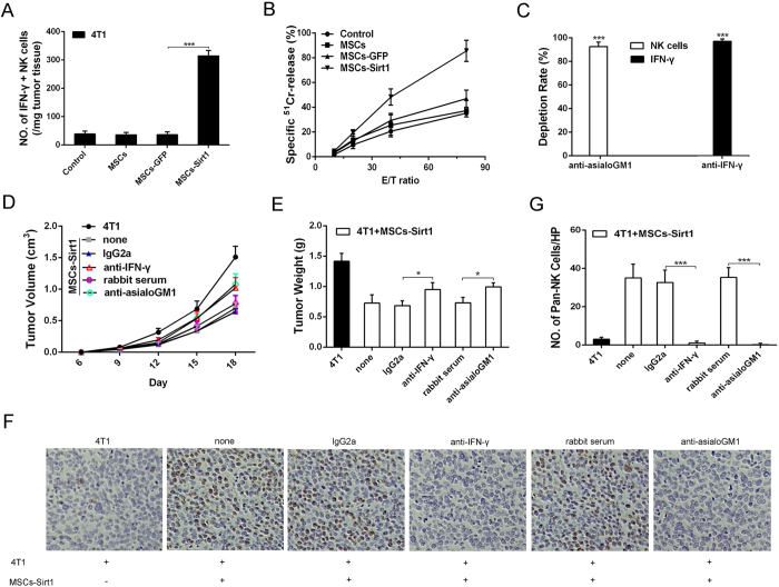
Figure 4. The effect of NK cells on tumor repression in tumor-bearing mice.
(A) Tumor infiltrating cells were isolated from the tumors of mice and were stained with antibody against NK cells, followed by intracelluar IFN-γ staining. (B) Splenocytes (effector, E) were isolated from the mice after sacrifice and assayed against YAC-1 cells (target cell, T). Cell lysis was determined in triplicate and 51Cr isotope release assay at different effector-to-target cell (E/T) ratios were performed to determine the cytotoxic potential of effector populations. (C) Mice were depleted of IFN-γ and NK cells by anti-IFN-γ mAb and rabbit anti-asialoGM1 antiserum by intraperitoneal injection, respectively. The depletion rates were also texted through ELISA kit and flow cytometry. Each group consists of 5 mice. (D) 4T1 tumor growth under IFN-γ or NK cell subset depletion was examined again. Mice treated with an irrelevant mouse monoclonal IgG2a or normal rabbit serum at the same dose were included as control. The width and length of 4T1 tumors were measured per 3 days, then the volume of tumor was calculated using the formula: volume = width2 × length × 0.5236. (E) Tumor weights were measured after they were harvested from the mice. (F) Tumor tissues were analyzed for immunohistochemical staining of CD49b for pan-NK cells (original magnification: × 200). (G) The number of pan-NK cells per HP (mean ± SD) was quantified. Each group consists of 5 mice. HP, high power field (400 × ). *, P < 0.05; ***, P < 0.001.

