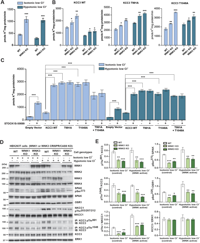
Figure 4. WNK3-SPAK regulates the phosphorylation and function of KCC3.
(A) 86Rb+ uptake assays in WNK3 WT and KO ES cells. WT and WNK3 KO cells60 were incubated with the indicated isotonic and hypotonic conditions (see Methods) for 30 min in the presence of 1 mM ouabain and 0.1 mM bumetanide. 86Rb+ uptake proceeded for 10 min and was quantified by scintillation counting. Results are presented as means ± SEM for triplicate samples. ***p < 0.001; **p < 0.01; *p < 0.05, when compared to WT values under the same conditions. (B) 86Rb+ uptake assays in WNK3 and WNK1 KO HEK293 cells. The indicated cells were transfected with constructs encoding a Flag empty vector or the indicated WT or mutant constructs (against KCC3 Thr991 and Thr1048) of N-terminal FLAG-tagged KCC3. 36 h post-transfection, cells were treated for 30 min with the indicated conditions and 86Rb+ uptake assays were then carried out in the presence of 1 mM ouabain and 0.1 mM bumetanide and quantitated by scintillation counting. Results are presented as in (A). Cell lysates from in parallel experiment were also subjected to immunoblot analysis (Supplementary Figure 4D,E). (C) 86Rb+ uptake assays in the presence of STOCK1S-50699. HEK293 cells were transfected and treated as in (B). 10 min 86Rb+ uptake assays were carried out in the presence of 1 mM ouabain and 0.1 mM bumetanide plus 10 μM of STOCK1S-50699 (indicated in the figure as +IN) and quantitated by scintillation counting. (D) HEK293T WT, WNK3 KO and WNK1 KO cells (see Methods) were treated for the indicated times with the indicated conditions. Harvested cell lysates were subjected immunoprecipitation (IP) and/or immunoblot (IB) with the indicated antibodies. (E) Graphs show quantitation of Western blot ratios (phospho-KCC3)/(total KCC3) (n = 3, means ± SD). ***p < 0.001; **p < 0.01; *p < 0.05; ns: non-significant (unpaired t-test). Under hypotonic low Cl− conditions, WNK1 KO cells and WNK3 KO cells both exhibited apparently decreased phosphorylation of heterologous KCC3 at Thr991 (p < 0.001) and Thr1048 (p < 0.001). WNK1 KO HEK293T cells and WNK3 KO HEK293T cells exhibited apparent decreases in phosphorylation of NKCC1 Thr203/Thr207/Thr212, SPAK Ser373, and OSR1 Ser325 (p < 0.05).

