
Fig. 1.
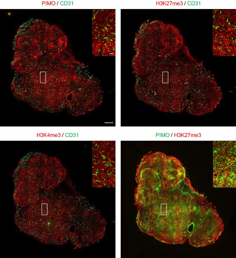
Tumor hypoxia increases trimethylation at H3K4 and H3K27. Representative images of whole tumor parallel sections stained for pimonidazole (hypoxia marker, red) and CD31 (PECAM-1; endothelial cell surface marker, green; upper left), H3K27me3 (red) and CD31 (green; upper right), H3K4me3 (red) and CD31 (green; lower left); scale bar represents 1 mm. The color-merged image (lower right) shows pimonidazole (green) and H3K27me3 (red). Captions show enlargements of the indicated areas; scale bar represents 100 µm