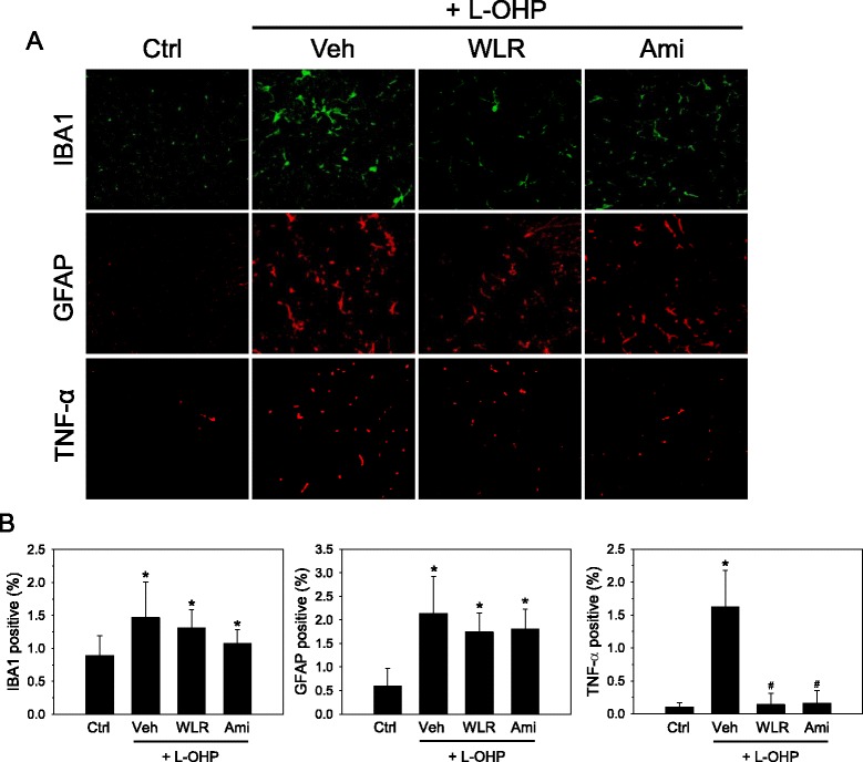
Fig. 3.

Anti-inflammatory effects of WLR in the spinal cords of the OXIPN animals. a Induction of neuropathy by oxaliplatin (L-OHP) and drug administration were done as described in Fig. 2. Four weeks after administration of WLR (250 mg/kg, 6 times a week) or amifostine (Ami, 100 mg/kg, once a week), the spinal cords of experimental animals were procured and subjected to immunohistochemical analyses. Microglias (green) and astrocytes (red) in the lumbar dorsal horn of the mouse spinal cords (L3-L4) were detected with IBA1 and GFAP specific antibodies, respectively (X200). Activated microglias and astrocytes showed marked hypertrophy in the oxaliplatin-treated animals. b Activated astrocytes and microglial cells were quantified by measuring the corresponding IBA1 and GFAP immunoreactivities, respectively, and were expressed as a percentage of immune-positive areas per total image areas. *p < 0.05 vs saline control mice (Ctrl); # p < 0.05 vs. oxaliplatin (L-OHP) and vehicle (Veh)-treated mice. The data represent the combined results of n = 5 (IBA1 and GFAP) or n = 4 (TNF-α) experiments
