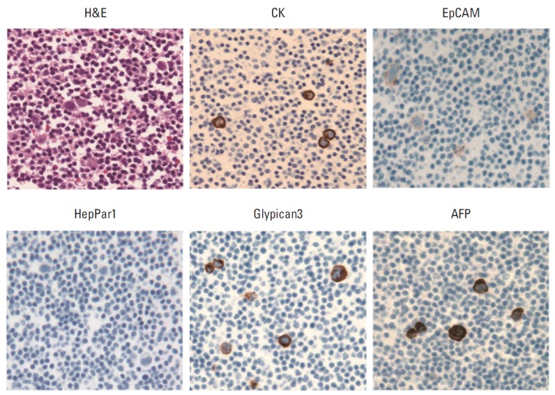
Fig. 2.

Histologic and immunohistochemical images (×400) of peripheral blood cell block containing spiked hepatocellular carcinoma cell line HepG2. The HepG2 cells show strong expression of cytokeratin (CK), Glypican3, and α-fetoprotein (AFP), and weak expression of epithelial cell adhesion molecule (EpCAM), but the cells are negative for HepPar1.
