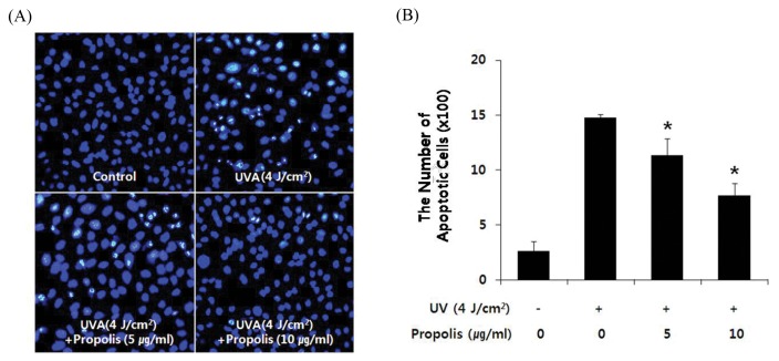
Fig. 1.
Propolis inhibited UVA-induced apoptosis in HaCaT cells. Cells were pretreated with or without propolis (5 and 10 μg/mL) for 1 hr and then irradiated with UVA (4 J/cm2). After incubation for 6 hr, cells were stained with Hoechst 33258. (A) Representative morphology visualized under a fluorescence microscope (BX70, Olympus, Tokyo, Japan). Cells with brightly fluorescent and fragmented nuclei were apoptotic. (B) Quantitative analysis of the percentage of apoptotic cells. The percentage of apoptotic cells = The numbers of apoptotic cells/(The numbers of apoptotic cells + The numbers of viable cells). *P < 0.05 when compared to control group.

