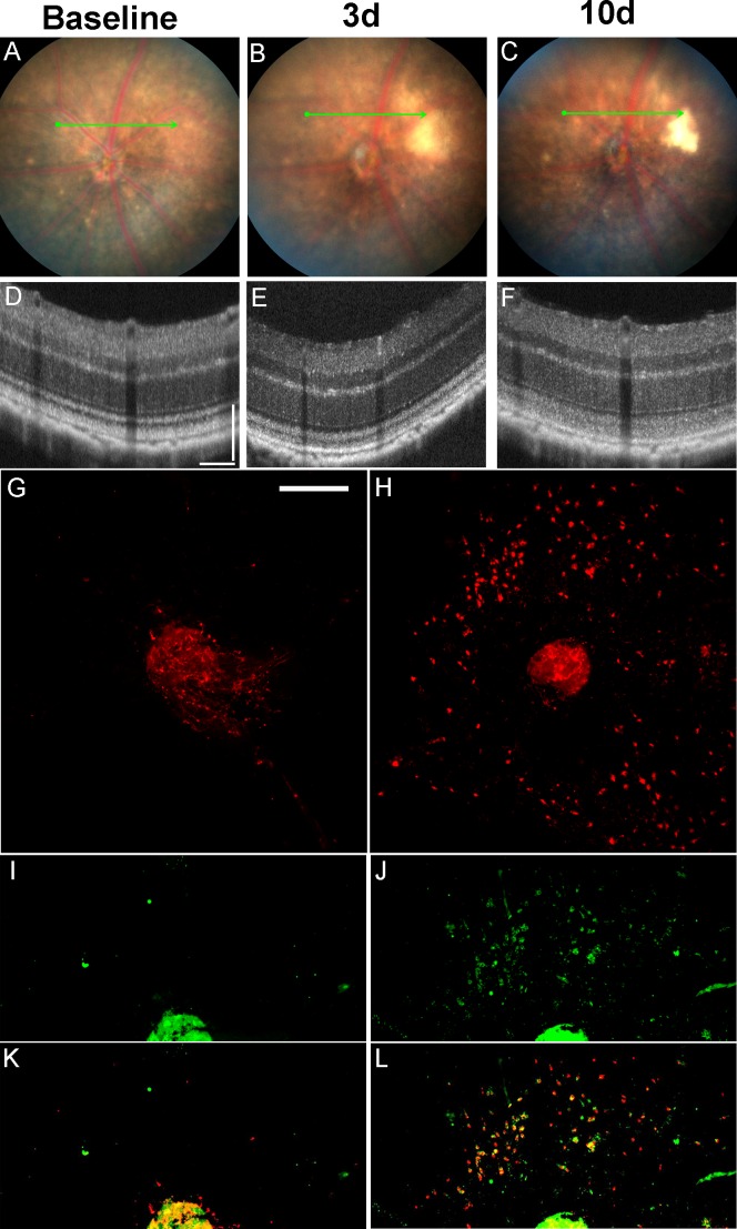
Figure 3.
Fundus photos, OCT and RPE flat-mounts of eyes treated with a “Light-only” FCD-LIRD protocol. (A–F) Fundus photos and OCT images of a C57BL/6J eye at baseline (A, D) and also 3 d (B, E) and 10 d (C, F) after exposure to 125 K lux of white light for 30 minutes. (G–L) RPE flat-mounts of two C57BL/6J eyes stained with Iba-1 (G, H), CD16 (I, J), or merging of the two channels (K, L). Left corresponds to an untreated eye (G, I, K), while right (H, J, L) corresponds to an eye collected 8 days after treatment with 125 K lux of white light for 30 minutes using the FCD-LIRD protocol. Optical coherence tomography scale bars: 100 μm for vertical and horizontal bars. Flat mount scale bar: 250 μm.

