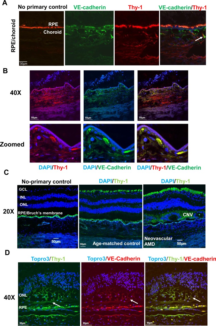
Figure 2.
Thy-1 is expressed at human choroids and CNV lesions. Immunostaining of Thy-1 (A) in retinal cryosections from a 25-year-old donor (red, Thy-1; green, VE-cadherin; autofluorescence of RPE shown in Alexa Fluor 568 and no primary control); (B) in paraffin-embedded sections from a 79-year-old donor with neovascular AMD (blue, 4′,6-diamidino-2-phenylindole [DAPI]; green, VE-cadherin; red, Thy-1); and (C, D) in paraffin-embedded sections from 75-year-old donor with neovascular AMD and an age-matched control donor without AMD (C) (blue, DAPI; green, Thy-1; magnification 20×) and at VE-cadherin–labeled CNV lesions (D) (green, Thy-1; red, VE-cadherin; blue, TO-PRO3, magnification, 40×; arrows point to CNV lesion).

