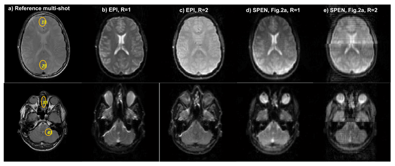
Figure 6.
Single-slice brain images of a volunteer scanned at two different locations. (a) Reference spin echo multi-scan images. (b) Spin-echo EPI images for R=1. (c) Idem for R=2. (d) Single-band hybrid SPEN, processed by SR and FT. (e) Parallelized Hybrid SPEN collected using the sequence in Fig. 2a, R = 2, and processed by SR-SENSE. Common parameters: FOV = 22x22 mm2, slice thickness = 5 mm. Common R=1 SPEN and EPI scan parameters: number of acquired points 80x80, Tacq = 40ms; for EPI TE = 45ms; for SPEN TE = 5-68 ms, Gexc = 0.05 G/cm, Texc = 40ms. Common R=2 SPEN and EPI scan parameters: number of acquired points 80x40, Tacq=20ms; for EPI TE = 30 ms and for SPEN TE = 5-42ms, Gexc = 0.1 G/cm, Texc=20ms. Multi-scan parameters: number of acquired points 384x384, TE = 71ms, TR = 1500ms. Also shown are SNRR=2/SNRR=1 ratios measured for SPEN and EPI in the (1)-(4) regions displayed in (a).

