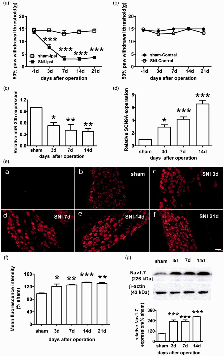
Figure 3.
Increased expression of Nav1.7 and decreased expression of miR-30b in the ipsilateral L4–6 DRG of SNI rats. (a, b) The paw withdrawal response to mechanical stimuli was evaluated in the sham and SNI (n = 6). The data are the mean ± SEM. ***P < 0.001 compared with the sham. (a) Responses of ipsilateral paws. (b) Responses of contralateral paws. (c) miR-30b expression levels in the DRG of the sham and SNI ipsilateral sides at 3, 7, and 14 days after the operation (n = 4). *P < 0.05, **P < 0.01 compared with the sham. (d) Expression level of SCN9A mRNA in the DRG (n = 4). *P < 0.05, **P < 0.01, ***P < 0.001 compared with the sham. (e) Representative immunofluorescence images of Nav1.7 expression in the DRG of the sham (b) and SNI ipsilateral sides at 3 (c), 7 (d), 14 (e) and 21 days (f) after the operation. (a) Section of DRG for negative control experiment, in which anti-Nav1.7antibodies was pre-incubated with Nav1.7 protein. Scale bar: 50 µm. (f) Quantification of Nav1.7 labelling intensity in (e) normalized to the sham values. n = 3. *P < 0.05, **P < 0.01, ***P < 0.001 compared with the sham. (g) Representative Western blot band and graph showing Nav1.7 expression in the DRG of the SNI ipsilateral sides (n = 4) at different time points after injury. β-actin served as a loading control. ***P < 0.001 compared with the sham.

