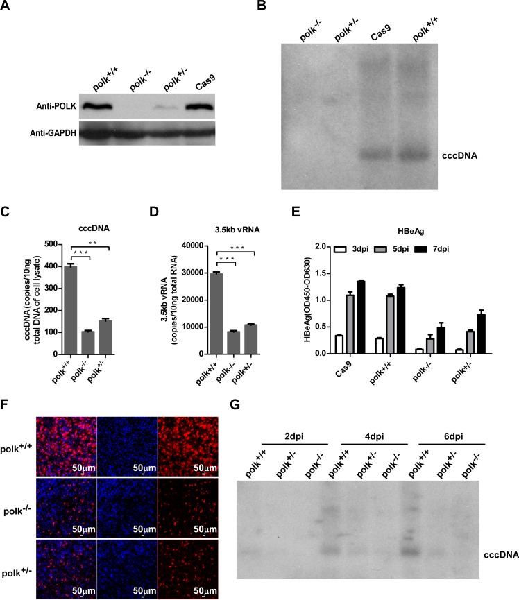
Fig 5. POLK is required for HBV cccDNA synthesis.
(A) POLK expression in parental HepG2-NTCP cells and HepG2-NTCP cell clones bearing disrupted polk gene in one (HepG2-NTCPpolk+/-) or both alleles (HepG2-NTCPpolk-/-) was determined by Western blot assay. GAPDH protein served as a loading control. (B-F) The indicated HepG2-NTCP-derived cell lines were infected with HBV. HBV cccDNA on 7 dpi was detected by Southern blot (B). The amounts of cccDNA (C) and 3.5kb vRNA (D) on 7 dpi were measured by qPCR assays. Data were analyzed by an unpaired two-tailed t test. ** p<0.01 and *** p<0.001. Secreted HBeAg from the indicated HepG2-NTCP cells on 3, 5, 7 dpi was measured by ELISA (E). HBcAg in the infected cultures was stained with 1C10 mcAb (red). Nuclei were stained with DAPI (blue) (F). Images were captured with a Nikon A1-R confocal microscopy, scale bars, 50 μm. (G) The timecourse of cccDNA synthesis in HBV infected HepG2-NTCP cells with intact or disrupted POLK gene was determined by Southern blot analysis.

