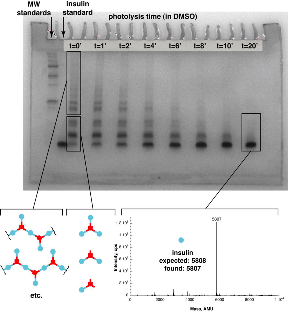
Figure 3. Gel analysis of insulin polymer and photolysis in DMSO.
Gel, Lane 1: Molecular weight standards ladder. Lane 2: insulin. Lane 3: Insulin polymer mixture (1:1:1 TD:IMA:IDA) at time 0. Lanes 4–10: Insulin polymer solution after photolysis of 1 min., 2 min., 4 min., 6 min., 8 min., 10 min., 20 min. using a 365nm Blak-Ray lamp. Mass spectrum of polymer photolysis product in DMSO (lower right): Deconvoluted ESI-MS of product formed after 20 minutes of photolysis of the insulin polymer. Expected mass for insulin: 5808. Observed: 5807.

