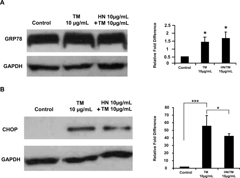
Fig 4. Increased expression of the ER stress markers GRP78 and CHOP by TM treatment of hRPE cells and the effect of HN.
Confluent hRPE cells were pretreated for 12 hours with or without 10 μg/mL HN. Cells were then treated with 10 μg/mL HN and/or 10 μg/mL TM for 12 hours. (A) Expression of GRP78 by Western blot analysis was significantly higher in TM and HN plus TM groups compared to control. (n = 4, *p < 0.05). (B) Expression of CHOP by Western blot analysis was significantly different between TM and HN plus TM groups compared to controls. However, treatment with HN along with TM reduced the expression of CHOP as compared to TM alone. Bar graph represents protein expression quantified by densitometry normalized to GAPDH. Data are mean ± SEM (n = 3). Asterisks represent ***p < 0.001, **p < 0.01. C- Control.

