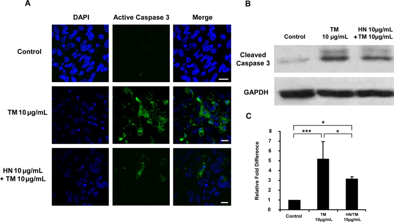
Fig 5. Activation of caspase 3 by TM-induced ER stress in hRPE cells and suppression of activated caspase 3 by HN.
Confluent hRPE cells were pretreated for 12 hours with or without 10 μg/mL HN. Cells were then treated with 10 μg/mL HN and/or 10 μg/mL TM for 12 hours. (A) Western blot analysis of total cell lysates probed with active caspase 3 antibody showed increased amounts of active caspase 3 with TM, and attenuation of active caspase 3 with HN. (B) Protein expression quantified by densitometry as shown as a ratio normalized with GAPDH. Data are mean ± SEM (n = 3). Asterisks represent *p<0.05, ***p<0.001.

