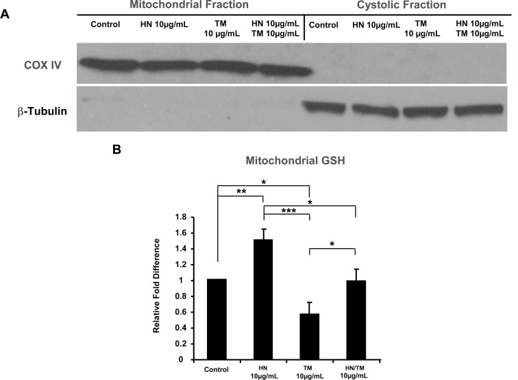
Fig 9. HN restores mitochondrial GSH from depleted levels due to ER stress.
Confluent hRPE cells were pretreated for 12 hours with or without 10 μg/mL HN. Cells were then treated with 10 μg/mL HN and/or 10 μg/mL TM for 12 hours. Cells were then fractionated into mitochondrial and cytosolic components using a mitochondrial/cytosol fractionation kit. (A) Western blots of COX IV (mitochondrial marker) and β-Tubulin (cytosolic marker) revealed there is negligible cross-contamination between the two fractions. (B) Mitochondrial GSH measurements showed decreased mitochondrial GSH with TM treatment, and repleted mitochondrial GSH with HN co-treatment. Data presented in the bar graph are mean +/- SD of 3 experiments. (*p<0.05, **p<0.01, ***p<0.001)

