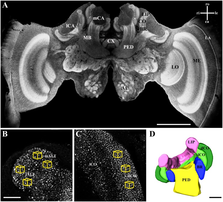
Fig 2. Synapsin immunostaining and 3D calyx reconstruction of a forager honeybee brain.
A Confocal image of a frontal section through the brain after whole-mount immunolabeling for synapsin. Calyx volume and MG density were quantified in one of the medial calyces (mCA). In the magnified view of the lip (B) and the collar (C), synapsin-labeled projection neuron boutons (MG) were counted in defined volumes (1000 μm3; yellow cubes) in three regions: mALT innervated lip, lALT innervated lip, and dense collar (dCO). D Cross section of the volume reconstruction of the mCA rendered from confocal image stacks. AL, antennal lobe; BR, basal ring; mCA, medial calyx; lCA, lateral calyx; CX, central complex; dCO, dense collar; lCO, loose collar; LA, lamina; LO, lobula; MB, mushroom body; ME, medulla; PED, peduncle. Axes: ca, caudal; le, left; ri, right; ro, rostral. Scale bar in A is 500 μm, in B (and C) 25 μm, and in D 100 μm.

