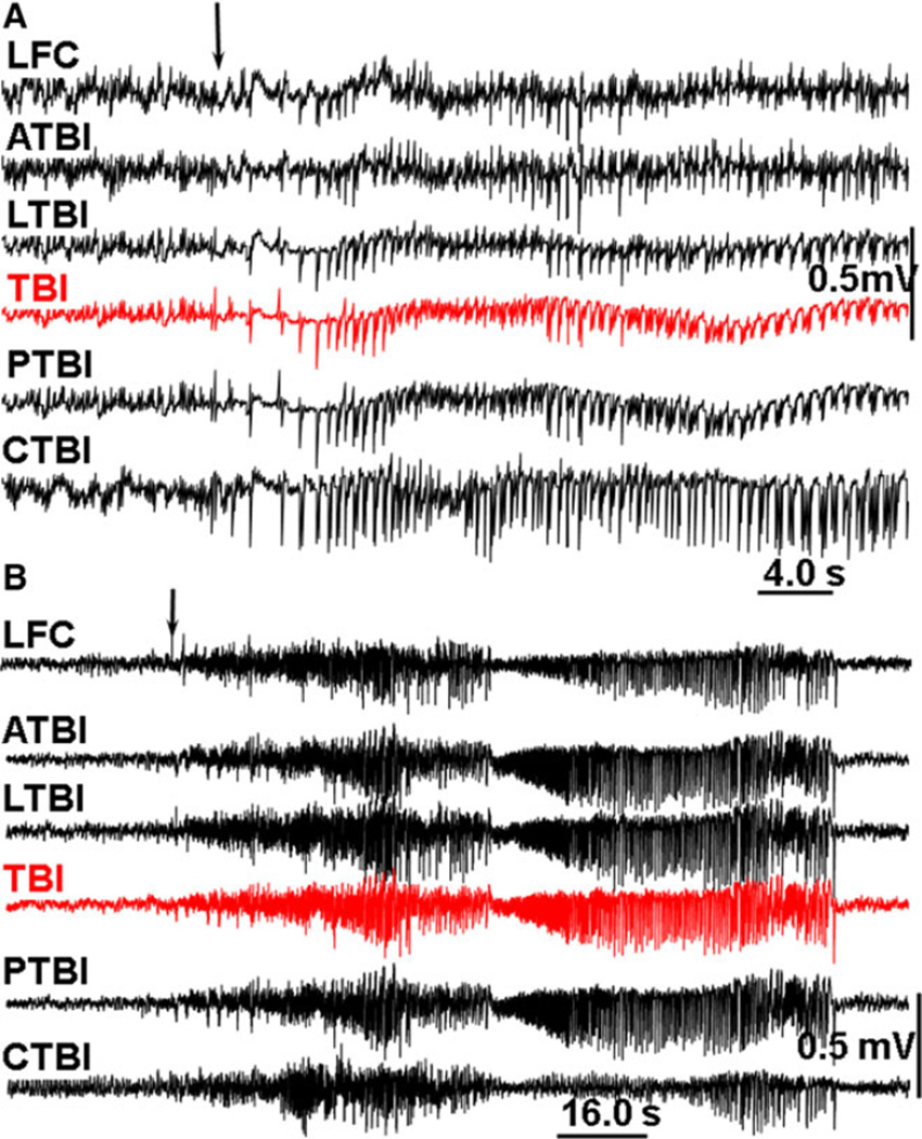
Figure 6.
(A) An example of spontaneous seizure on day 2 after TBI. (B) An example of seizure 167 days after FPI. LFC, and RFC: left and right frontal cortex; TBI, TBI core, place of application of FPI; ATBI, LTBI, PTBI and CTBI, correspond respectfully to areas of neocortex anterior, lateral, posterior, and contralateral to the TBI core. Epilepsia © ILAE

