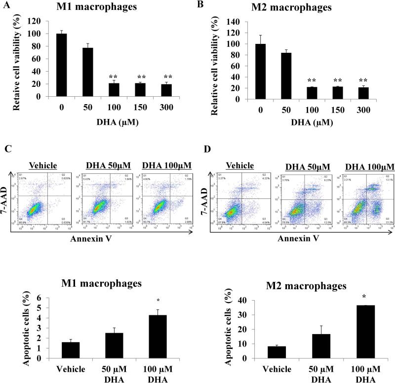
Figure 4.
Effects of different doses of docosahexaenoic acid (DHA) on M1 and M2 macrophage cell viability and apoptosis. THP-1 cells were treated with 320 nM phorbol 12-myristate 13-acetate (PMA; Sigma). After 24 h, PMA was removed and cells were treated with 10ng/ml lipopolysaccharide (LPS) for 3 h to get M1 macrophages. For THP-1 M2 macrophages differentiation, cells were treated with 320 nM PMA (Sigma). After 24 h, PMA was removed and cells were treated with 20 ng/mL interleukin (IL)-4, and 20 ng/mL IL-13 (Sigma, USA) for an additional 24 h. A. M1 and B. M2 macrophages were incubated with DHA overnight, cell viability was analyzed by MTS assay. C and D. M1 and M2 macrophages were incubated with DHA overnight, cells were collected and stained with Annexin V and 7-AAD, apoptosis was analyzed by flow cytometry. Data are presented as the mean ± SD (*, p<0.05; **, p<0.01).

