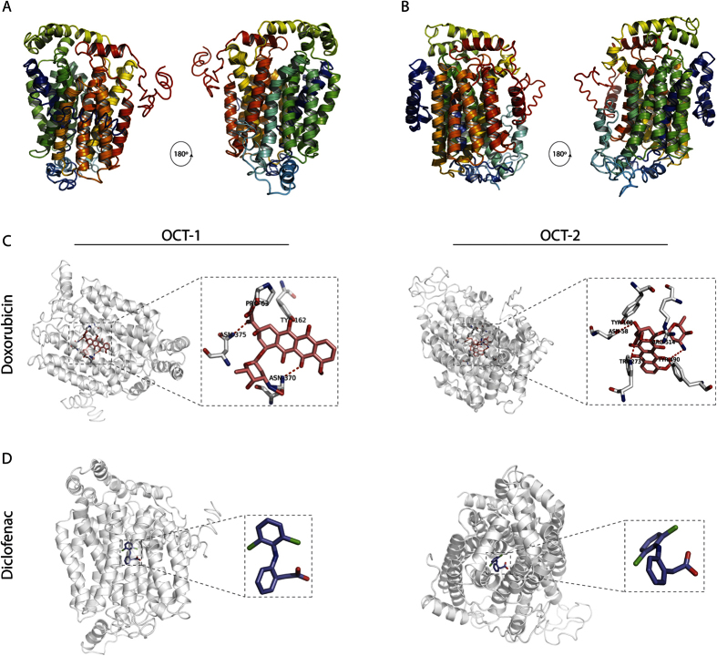
Figure 4. Structural modelling and protein-ligand docking of C. elegans OCT-1 and OCT-2.
(A,B) Structural models of OCT-1 and OCT-2 were generated on the basis of GLUT3 (PDB ID: 5c65) structure and sequence conservation. N-terminal and C-terminal are coloured blue and red, respectively. (C,D) Predicted binding models of OCT-1 and OCT-2 with cationic and anionic ligands doxorubicin (pink) and diclofenac (purple), respectively. Residues making polar contacts with the ligands are depicted with sticks and represented with dotted red lines; oxygen atoms are coloured in red, nitrogen atoms in blue and carbon atoms in white. All representative figures were rendered with PyMol.

