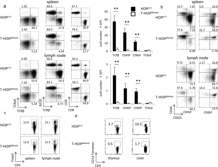
Figure 2. Marked decrease of CD4+ or CD8+ T cells in T-HOIPΔlinear mice.
(a) Spleen cells from T-HOIPΔlinear mice and HOIP+/+ mice were stained with anti-CD4, anti-CD8α, anti-TCRβ, anti-TCRγ and anti-B220 antibodies and the frequencies of cells expressing TCRβ/TCRγ, TCRβ/B220 and CD4/CD8 were evaluated by gating on lymphocytes in an FSC/SSC gate. The number indicates the percentage of each population within the viable population (left and middle panels) and the percentage of each population in the TCRβ+ population (right panel). Data show absolute numbers of total thymocytes, TCRβ+ cells, TCRγ+, CD4+CD8− (CD4SP) and CD4−CD8+ (CD8SP) cells from T-HOIPΔlinear (open) or HOIP+/+ (filled) mice at the age of 8 weeks. Data are presented as means ± SEM. **p < 0.01. Spleen cells or liver lymphocytes from T-HOIPΔlinear or HOIP+/+ mice were stained with a combination of (b) anti-CD4, anti-CD8α, anti-CD44 and anti-CD62L antibodies, or (c) anti-CD4, anti-TCRβ and anti-Foxp3 or (d) anti-CD4 and anti-CD1d tetramer. The frequency of CD44/CD62L cells was evaluated by flow cytometry by gating on CD4+CD8− (CD4SP) or CD4−CD8+ (CD8SP), or CD4/Foxp3 using a primary FSC/SSC gate to identify lymphocytes expressing CD4/CD1d. The number indicates the percentage of each population within the viable population. The data in these figures are representative of four independent experiments.

