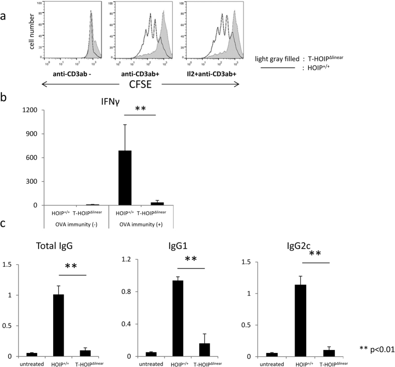
Figure 3. Impaired proliferation and cytokine secretion of CD4+ T cells in T-HOIPΔlinear mice.
(a) CFSE-labelled CD4+ T cells from spleens of control (line) or T-HOIPΔlinear (filled gray) mice were stimulated for 3 days on plates coated with anti-CD3 mAb (1 μg/mL) in the absence or presence of recombinant IL-2 (10 U/mL). HOIP+/+ or T-HOIPΔlinear mice at the age of 8 weeks were immunized by OVA protein (10 μg/mL) emulsified in CFA. (b) Serum IFN-γ was evaluated by ELISA ten days after immunization. Data show means ± SEM. **p < 0.01. (c) Serum anti-OVA IgG, IgG1 or IgG2c levels were evaluated by ELISA ten days after immunization. Data show means ± SEM. **p < 0.01. The data in these figures are representative of four independent experiments.

