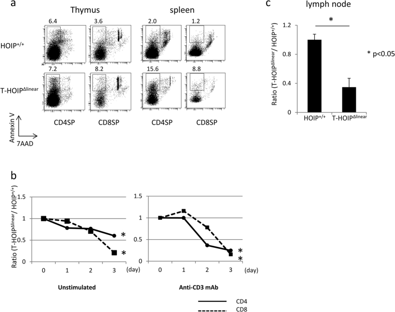
Figure 5. Impaired survival of T cells in T-HOIPΔlinear mice.
(a) Thymocytes and spleen cells were stained with anti-CD4 and anti-CD8 antibodies together with Annexin V and 7AAD. CD4+ or CD8+ T cells with an Annexin V+7AAD− phenotype were evaluated. The number indicates the percentage of each population. (b) Isolated CD4+ or CD8+ T cells from HOIP+/+ or T-HOIPΔlinear mice were cultured in the absence or presence of plate-coated anti-CD3 mAb. The cell number after the indicated number of days was counted. The value is calculated from the number of T-HOIPΔlinear/number of HOIP+/+ cells. *p < 0.05. (c) Isolated CD4+ T cells from HOIP+/+ (CD45.2) or T-HOIPΔlinear mice (CD45.2) were transferred into nonirradiated B6 mice (CD45.1). The number of donor cells 7 days after transfer was counted. Data are shown as means ± SEM. *p < 0.05. The data in these figures are representative of five independent experiments.

