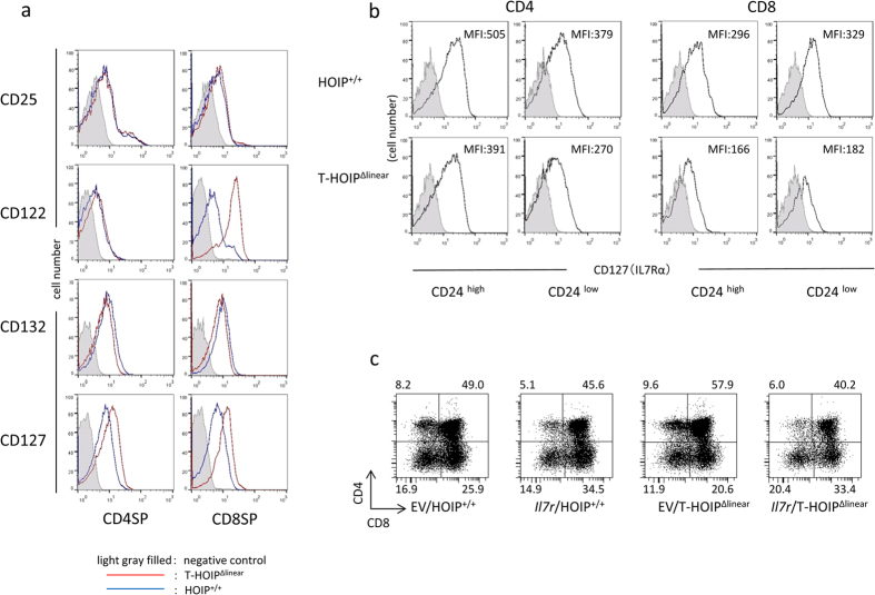
Figure 6. Defective IL-7Rα in thymocytes of T-HOIPΔlinear mice.
(a) Spleen cells from T-HOIPΔlinear (red) or HOIP+/+ (black) mice were stained with anti-CD4, anti-CD8α, anti-CD25, anti-CD122, anti-CD127 and anti-CD132 antibodies. The expression of CD25, CD122, CD127 and CD132 in CD4+CD8− (CD4SP) or CD4−CD8+ (CD8SP) was evaluated by flow cytometry. The negative control cells were stained with isotype controls (filled gray). (b) Thymocytes from T-HOIPΔlinear or HOIP+/+ mice were stained with anti-CD4, anti-CD8α, anti-CD24 and anti-CD127 antibodies. The expression of CD127 by CD4+CD8−CD24hi or CD4+CD8−CD24low, CD4−CD8+CD24low or CD4−CD8+CD24hi cells was evaluated by flow cytometry. As the negative control, cells were stained with isotype controls (filled gray). The number indicates the mean fluorescence intensity (MFI) of each population in the viable population. (c) Fetal thymocytes (day 15 fetal age) were infected with control retrovirus (EV) or retrovirus containing the CD127 gene (Il7r) and cultured in dGu-treated fetal thymus for 7 days. Thymocytes were stained with anti-CD4 and anti-CD8α antibodies and the expression gated on GFP+ cells was evaluated by flow cytometry. The number indicates the percentage of each population in the viable population. The data in these figures are representative of four independent experiments.

