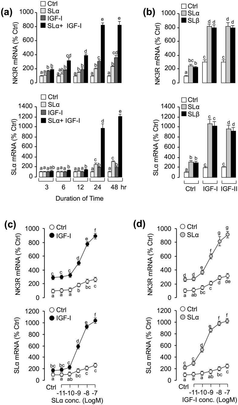
Figure 3. Functional interaction between SL and IGF on the regulation of NK3R and SLα gene expression.
(a) Time course of carp SLα, IGF-I and SLα+ IGF-I on NK3R and SLα mRNA expression. In this experiment, carp pituitary cells were incubated from 3 to 48 hr with SLα (30 nM) alone, IGF-I (50 nM) alone and SLα (30 nM)+ IGF-I (50 nM). (b) Synergistic effects of SLα/β and IGF-I/-II on the simulation of NK3R and SLα mRNA expression. In this case, carp pituitary cells were challenged for 24-hr with SLαor SLβ (30 nM) co-treatment with either IGF-I or IGF-II (50 nM). (c) Effect of SLα (0.01–100 nM) treatment on basal and IGF-I (50 nM)-induced NK3R and SLα mRNA expression in carp pituitary cells. (d) Effects of IGF-I (0.01–100 nM) treatment on basal and SLα(30 nM)-induced NK3R and SLα transcript levels in carp pituitary cells. After drug treatment, total RNA was isolated for real-time PCR of NK3R and SLα mRNA. In the data present (mean ± SEM), the groups denoted by different letters represent a significant difference at p < 0.05 (ANOVA followed by Dunnett’s test).

